How and Why Do Brain Cells Die?
Sometimes our brain cells die, and researchers want to know why. Johns Hopkins University researchers may have found the answer.
By Norman Rusin
Chaining Proteins May Free Brain Cells from Disease
How did the king of Corinth, Sisyphus, outwit the god of death, Thanatos? By using the god’s own chains. When it was Sisyphus’s time to die, Zeus ordered Thanatos to chain Sisyphus up in Tartarus, the kingdom of the dead. King Sisyphus slyly asked Thanatos to demonstrate how the chains worked. As Thanatos was granting him his wish, Sisyphus seized the opportunity and trapped Thanatos in the chains instead. Once the god of death was bound by the strong chains, no one died.
Nowadays, another chain of events leads our brain cells to death: it is called parthanatos—keeping in its name the Greek myth’s memory—and it is responsible for injuries such as stroke and illnesses such as Alzheimer’s, Parkinson’s, and Huntington’s. However, Johns Hopkins University researchers have found the protein at the end of that chain of events that delivers the fatal strike by carving up a cell’s DNA. Their discovery may lead to new drugs that would prevent, stop, or weaken the process.
A Programmed Death
Conducted on laboratory-grown cells, these new experiments build on earlier work by research partners Ted Dawson, MD, PhD, now director of the Institute for Cell Engineering at the Johns Hopkins University School of Medicine, and Valina Dawson, PhD, professor of neurology. Their research groups found that—despite very different causes and symptoms—injury, stroke, Alzheimer’s disease, Parkinson’s disease, and the rare, fatal genetic disorder Huntington’s disease have a shared form of “programmed” brain cell death. The team looked closely at a protein called mitochondrial Apoptosis-Inducing Factor (AIF), a specialized part of a cell outside its nucleus that causes the cell’s programmed death. They knew that when AIF leaves its usual place in the energy-producing mitochondria of the cell and moves to the nucleus, it sparks the carving up of the DNA housed in the nucleus and leads to cell death.
“I can’t overemphasize what an important form of cell death it is; it plays a role in almost all forms of cellular injury,” Ted Dawson says. The research groups have spent years delineating each of the links in the parthanatos chain of events and the roles of the proteins involved. The current study, they say, has completed the chain.
RELATED: Neuromodulation: How We Manipulate Brain Cells
It Takes Two to Kill a Cell
Since AIF itself cannot cut DNA, it was necessary to look for proteins that interact with DNA. Yingfei Wang, assistant professor at the University of Texas Southwestern Medical Center, used a protein chip to screen thousands of human proteins to find such a protein. First, she dug out 160 proteins qualified for this task. Then, she used custom molecules, called small interfering RNAs, to stop each of those proteins’ manufacture, one by one, in lab-grown human cells to see if doing so would prevent cell death. One of the 160 proteins, known as macrophage Migration Inhibitory Factor (MIF), was a winner. “We found that AIF binds to MIF and carries it into the nucleus, where MIF chops up DNA,” Ted Dawson reports. “We think that’s the final execution step in parthanatos.”
Where Does MIF Strike?
Dawson cautions that while parthanatos is known to cause cell death in many brain conditions, MIF’s ability to chop up DNA has so far been definitively linked only with stroke—when the MIF gene was disabled in mice, the damage caused by a stroke was dramatically reduced. “We’re interested in finding out whether MIF is also involved in Parkinson’s, Alzheimer’s, and other neurodegenerative diseases,” he says, and that if so, and if an inhibitor of MIF proves successful in testing, it could have implications for treating many conditions.
The research group reports that in work to be published they have also identified a few chemical compounds that block MIF’s action in the lab-grown cells, protecting them from parthanatos. Ted Dawson says the group plans to test these compounds in animals and modify them to maximize their safety and effectiveness.
References
http://www.newswise.com/articles/brain-cell-executioner-identified
http://www.ninds.nih.gov/disorders/brain_basics/ninds_neuron.htm
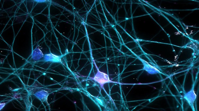
Featured image: Artist’s representation of neurons. Published on Flickr by Ardy Rahman.
—Norman is a freelance copy editor and journalist who assists writers to produce sound and telling communications. He graduated from the University of Pennsylvania with a PhD in Italian Studies. In his research, he looks at how science, literature, and art complement each other in our understanding of the world. Connect with him at normanrusin77@gmail.com.